
The history of the trochlear dysplasia in patella dislocation
175
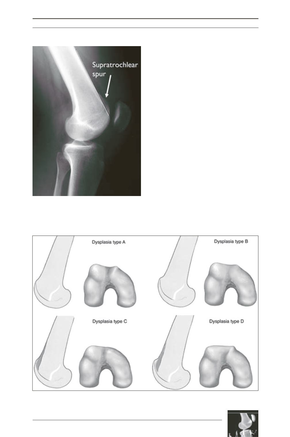
Type B
is pathologic for a flat trochlea with a
prominence and the additional
supratrochlear
spur
. In
Type C
, the trochlea is convex and has
the “crossing sign” and the “double contour
sign” on sagittal X-rays. In the most severe
Type D
, the trochlear groove is elevated above
the anterior femoral cortex; it has a hypoplastic
medial facet and a “cliff-pattern” appearance
with all three previous signs on the lateral
X-rays. This new classification scheme was
more reproducible and had a high observer
agreement [44, 45].
The interest on studying the morphology of
trochlear dysplasia on axial views was also
strong in a study by R. Biedert, who analyzed
the decreased trochlear depth caused by either
an elevated trochlea floor or a flattened lateral/
medial condylar height on MRI [46]. He
compared the height of the medial, central and
lateral third of the trochlea according to the
width of the lateral condyle and he discovered
that a reduced height of the lateral condyle
more than 77% was pathologic and that in more
Fig. 9: The supratrochlear spur on a true lateral
X-ray represents the prominence of the whole
trochlea.
Fig. 10: The four-type classification for trochlear dysplasia proposed by D. Dejour.


