
S. Lustig, C. Scholes, M. Coolican, D. Parker
144
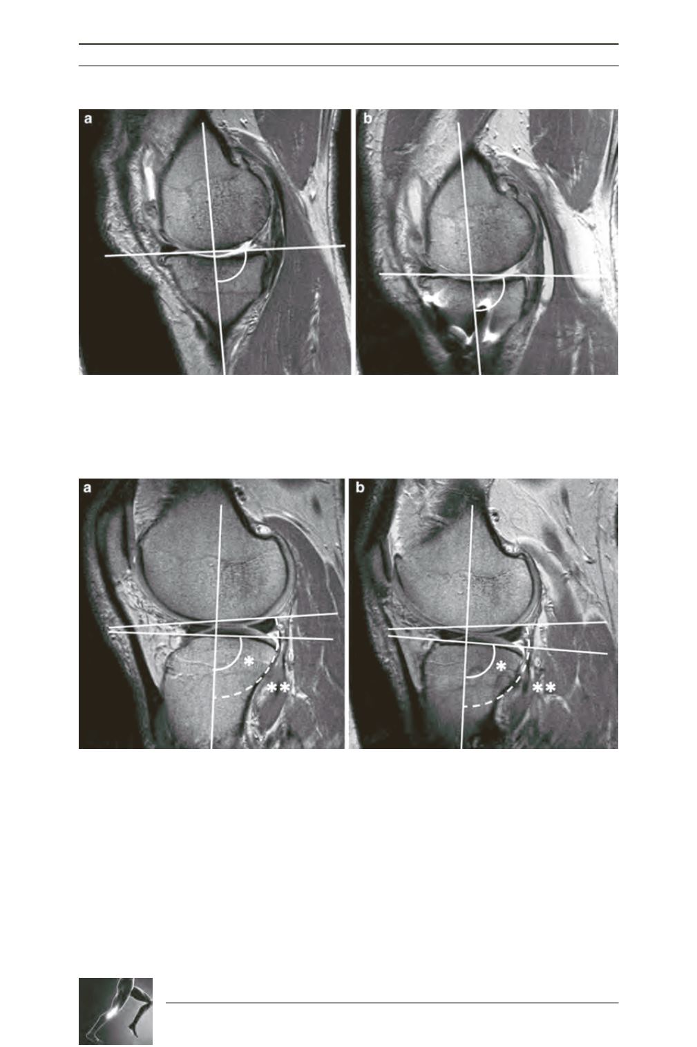
Fig. 2: Sagittal plane section in the middle of the medial tibial plateau was used for measurement of tibial
slope preoperatively (a) and postoperatively (b). The most superior points in the anterior and posterior part
of the medial tibial plateau were joined to obtain the line of the bony slope. It was not possible to identify
the limits of the medial meniscus due to osteoarthritic changes.
Fig. 3: Mid-sagittal images used for measurement of lateral tibial slope preoperatively (a) and post-
operatively (b). The most superior points in the anterior and posterior part of the lateral tibial plateau were
joined to obtain the line of the bony slope in the lateral compartment (single asterisk). Similarly, the highest
points of the interior and posterior horn of the lateral menisci were joined to generate the line of soft tissue
slope in the lateral compartment (double asterisks).


