
143
Introduction
In contrast to radiographic measurements, MRI
provides multiple slices of the knee joint in the
sagittal plane, making it possible to assess the
medial and lateral tibial slope separately. The
purpose of this study is to investigate the effect
of medial open-wedge high tibial osteotomy
(MOWHTO) on bony and meniscal slope in
the medial and lateral tibiofemoral com
partments. It was hypothesised that greater
changes on the medial tibial plateau would be
observed compared with the lateral one.
Methods
(fig. 1, 2 and 3)
A retrospective analysis of prospectively
collected data was performed on pre- and post-
operative MRIs from 21 patients (17 men and 4
women; age 52±9 years). Inclusion criteria
were varus alignment, medial compartment
osteoarthritis and election for a primary
MOWHTO. Each patient had a preoperative
and a post-operative highresolution MRI
(3Tesla, Magnetom Trio, Siemens AG) at an
average follow-up of 2.1 years. A previously
published method was used to measure bony
and meniscal slope for each compartment. The
difference between pre- and postoperative
tibial slope for both compartments was
calculated and associated with the amount of
frontal correction.
Cartilage, tibial slope
and HTO
S. Lustig, C. Scholes, M. Coolican, D. Parker
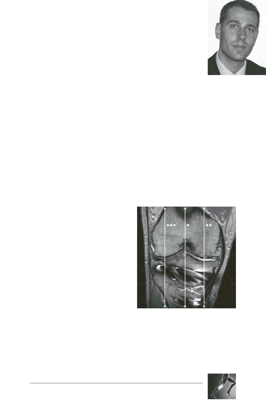
Fig. 1: Post-operativeMRI, 2 years after an opening-
wedge high tibial osteotomy. Sagittal images were
identified from the axial images at the joint line for
the mid-sagittal slice (single asterisk), the mid-
medial tibial plateau (double asterisks) and the
mid-lateral tibial plateau (triple asterisks).


