
Robotic Surgery: experience with Unicondylar Knee Arthroplasty
45
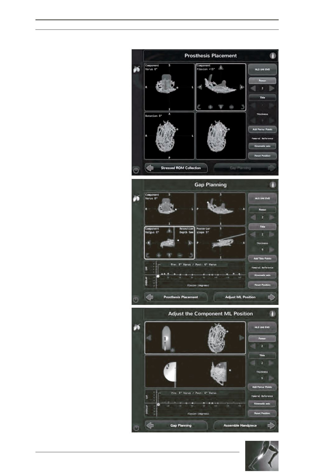
Fig. 3 : (A) The planning stage screen
shows where the user can adjust the
implant size and move the position of
the implant in all three planes to best
match the patient’s condyle. (B) The
gap planning screen shows the position
of the implant on the patient’s
condylarsurface. The graph at the
bottom of the screen illustrates the
virtual gap balance through a range of
motion predicted from implementing
the planned implant position and
tensioning the ligaments. (C) Contact
point screen, illustrates the contact
points on both the tibial and femoral
component as the knee goes through a
range of flexion.
A
B
C


