
S. Lustig, P. Neyret
46
Bone preparation is performed per the
manufacturer’s recommended technique for
robotic UKA with the Tornier HLS UNI
Evolution implants. The femoral component,
with a central lug and keel, is impacted rigidly
onto the prepared bone surface and the slotted
trough and peg hole on the femoral condyle
optimized positioning of the component. The
tibial implant in this particular design is a
cemented unconstrained all-polyethylene insert.
This implant design has reported good clinical
and radiological results [2]. It was designed
without lugs or keel to allowvariable positioning
on the AP axis based on intraoperative
assessment of positioning relative to the femoral
component. Once the gap balance through a
range of motion is checked with the trials
(fig. 5). both components are cemented (fig. 6).
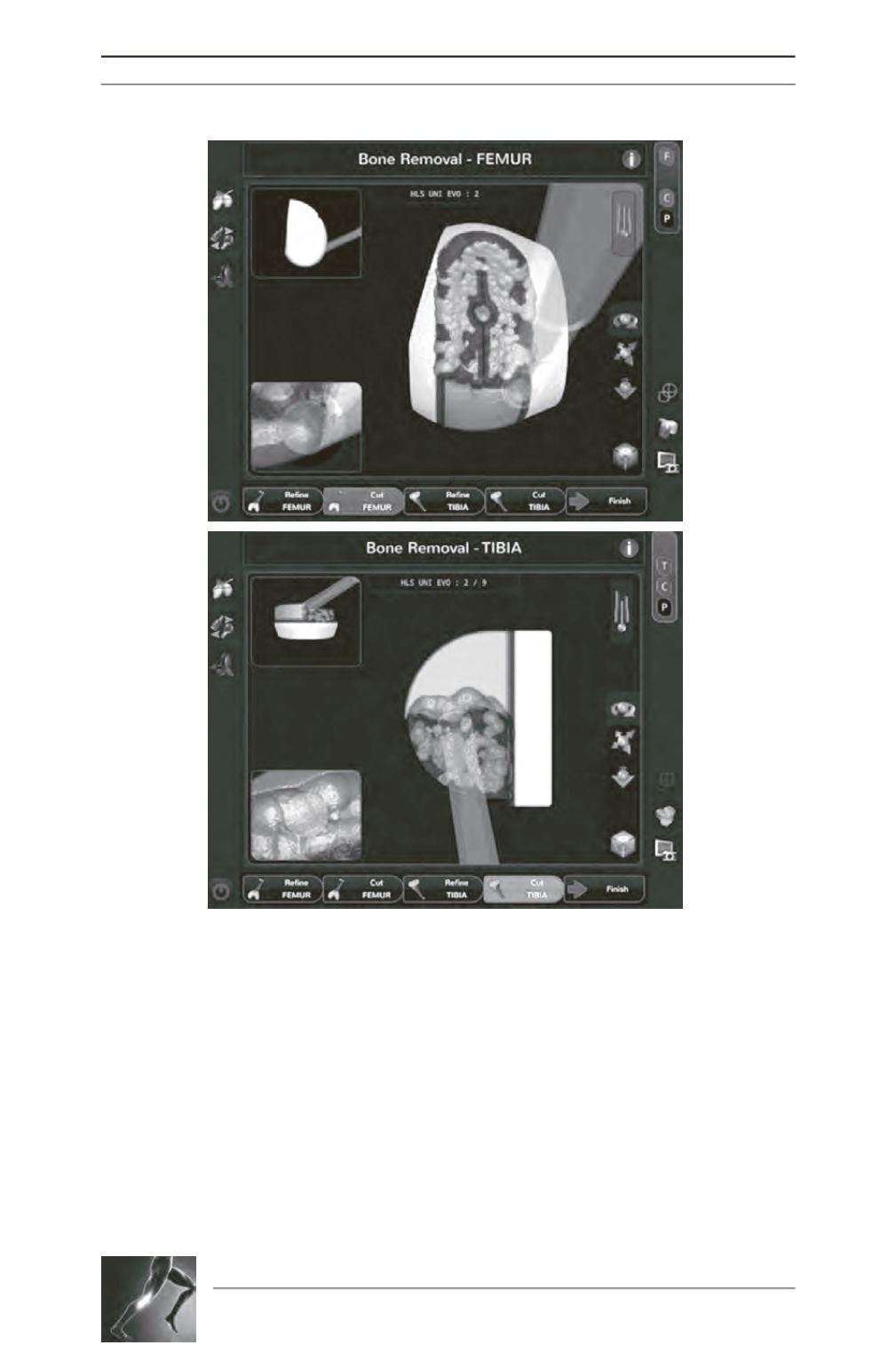
Fig. 4 : (A) Femur and (B) tibia cutting screens show midcutting. The
yellow surface is the “target” surface, green surface indicates 1mm of
bone still to be removed, blue surface indicates 2mm of bone still to be
removed, and the purple surface indicates 3mm or more bone still to be
removed.
A
B


