
MEDIAL SIDE
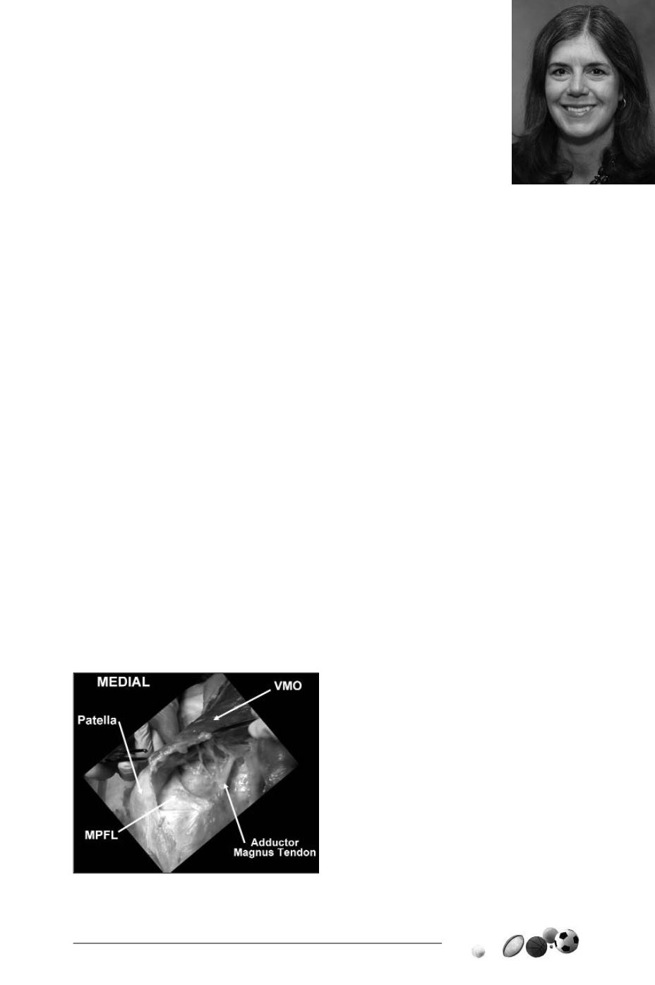
The Medial PF Ligament (MPFL) attaches
to the femur 10mm proximal and 2mm
posterior to the medial epicondyle, in the
saddle between the medial epicondyle and
the adductor tubercle (fig. 1). Its patella
attachment is approximated at the junction
of the upper and middle thirds of the patella,
typically at the location where the perimeter
of the patella becomes more vertical. It is the
prime soft tissue restraint to lateral patella
displacement. However, it is only significant
in early flexion.
As the knee progresses in flexion, trochlear
geometry, patellofemoral congruence and in
particular the
slope angle of the lateral
wall of the trochlea
provide the major
restraints to lateral patella displacement [8].
In trochlear dysplasia, the groove is often not
only flattened, but shortened. The shortened
groove combined with a high riding patella
(patella alta) will create a larger arc of
motion before the patella is protected by the
confines of the lateral trochlear wall.
MEDIAL SIDE VMO
ANATOMY
The MPFL is “covered” by the vastus media-
lis obliques (VMO) fibers. Typically, 35% of
the VMO is covered by these fibers in normal
anatomy [11]. With VMO dysplasia, the liga-
ment is “uncovered” most notably due to loss
of fiber obliquity (fig. 2).
37
ANATOMY AND BIOMECHANICS
OF PATELLOFEMORAL RESTRAINTS
(with particular reference to surgical concerns)
E. ARENDT
Fig. 1


