
DISCUSSION
Medial UKAs were performed for patients
with varus malalignement for any etiology
(arthritis or necrosis). As recommended in the
literature [5, 20, 21], we tried to conserve a
slight hypocorrection. However, some authors
[11, 18] tend to avoid hypocorrection because
of a risk of early wear. We did not have wear
related to that etiology. We found a slight val-
gus in the lateral UKA but it was not systema-
tic with some tibial implant measured in varus.
It may be explained by the origin of the defor-
mity: varus deformity tends to be mainly tibial,
but valgus deformity is usually mixed. It is
therefore important to perform a tibial cut at
nearly 90° in case of lateral UKA because the
surgeon does not want to perform a cut in val-
gus for a lateral UKA [21].
The mean tibial slope averaged 5°. For an
UKA we attempt to reproduce the tibial slope
of each individual. Hernigou
et al.
[10] sho-
wed the tibial slope had to be under 7°.
Beyond this point, there was a greater risk of
failure by increasing the anterior tibial transla-
tion and further rupture of the anterior crucia-
te ligament. Whiteside and Amador [25]
recommended a tibial slope between 3° and 7°,
whereas Matsuda
et al.
[13] found a tibial
slope averaging 10.7° for the medial tibial pla-
teau and 7.2° for the lateral tibial plateau.
Dejour and Bonnin [6] measured a mean
radiological tibial slope of 10° in the normal
population, but they highlighted also that a 10°
increase of the tibial slope would lead to
3.5mm increase of the anterior tibial transla-
tion. That increase could lead to rupture of the
anterior cruciate ligament and to greater bio-
mechanical constraint of the posterior part of
tibial component.
Regarding our rotation measurements, we may
point out the choice of the landmarks refe-
rences. For total knee arthroplasty, the tibial
component rotation has been analyzed several
times [22, 23]. At the proximal tibial, many
references have been used: the transepicondy-
lar axis, the medial border of the anterior tibial
tubercle, the center part of the patellar tendon,
the tibial insertion of the posterior cruciate
ligament (PCL), the medial and lateral parts of
the tibial plateaus and the posterior parts of the
tibial plateaus. Our choice (posterior parts of
the tibial plateaus) from Yoshioka [27] may be
criticized. Nevertheless there is a high variabi-
lity with the PCL tibial insertion [22] which is
also difficult to locate on a CT scan. The UKA
implant positioning in rotation is still challen-
ging and there is little scientific support to
assess the proper position. The tibial implant
rotation is guided by the antero-posterior cut.
It is sometimes said that the cut should be done
with the knee flexed at 90° and that the sur-
geon should aim the saw towards the hip. We
may accept the approximate feature of that cut.
In our series, the tibial implant was externally
rotated for both medial and lateral UKA with
no involvement of the femoral valgus. We
found only one study with analysis of the tibial
plateau positioning on the axial plane.
Campbell
et al.
[4] report great variation of the
tibial component orientation but he did not
give its entire data for the rotation in his
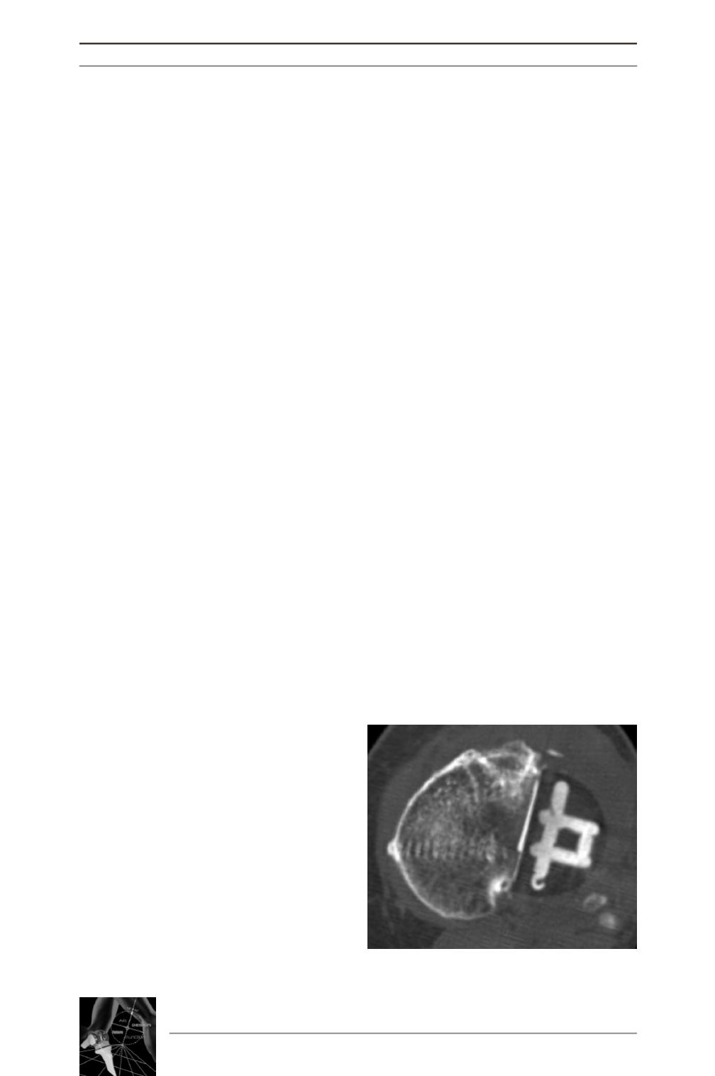
article. At last the CT-scan allowed to analyze
the tibial implant positioning on the axial
plane and may be to highlight a rotation mal-
positioning (fig. 3). The biomechanical conse-
quences of such malpositioning have never
been studied and we may find out a hidden
etiology of ongoing pain or early failure of an
UKA.
14
es
JOURNÉES LYONNAISES DE CHIRURGIE DU GENOU
176
Fig. 3: Lateral UKA with an excessive rota-
tion of the tibial plateau.


