


 195 / 324

DISCUSSION
The kinematics of the Journey CR knee
implant are on average comparable to the kine-
matic pattern of the native knee in these tests.
Apparently the joint surfaces of the anatomic
knee design with a dished medial insert surfa-
ce and a convex lateral insert surface and a
3 degrees varus of the joint line is guiding the
motion towards that of a normal knee joint. We
feel that correct balancing of the PCL during
implantation is of major importance in achie-
ving these results. The step-off guided spacer
technique to balance the PCL seems to work
well in this experiment, no lift off of the tibia
inserts occurred during trail implantation. The
preparation of the PCL with preservation of
the bony insertion was also safe since no liga-
ment ruptures occurred during the tests.
It is clear that the PCL is the main and largest
ligament structure in the flexion gap, and it
dictates the position of the contact point of
femur on tibia. The precise preparation of the
gaps is also important in achieving correct
kinematics. In two out of eight cases we nee-
ded to increase the slope of the tibia with 2-
3 degrees, thereby increasing the flexion gap
with 2-3mm. The described technique therefo-
re can be used easily.
We did not not use a PCL release in the tight
knees in this experiment since effects of
releases on roll-back and contact points were
not available in the literature and thus uncer-
tain. Moreover we feel that in case of a tight
flexion gap the PCL is not the cause of the pro-
blem but the gaps are not correct. As Christen
et al.
[3] have shown a 1-2mm flexiongap
increase causes a contactpoint change of 2-
4mm with the tibia moving anterior. This
explains why a limited increase of the tibia
slope with 2-3 degrees was sufficient to cor-
rectly balance the flexiongap and create a cor-
rect contact point.
In summary the kinematics of the anatomic
Journey CR total knee design seem to be simi-
lar to the kinematics of the native knee using a
measured bone resection technique and a spa-
cer guided PCL balancing.
14
es
JOURNÉES LYONNAISES DE CHIRURGIE DU GENOU
194
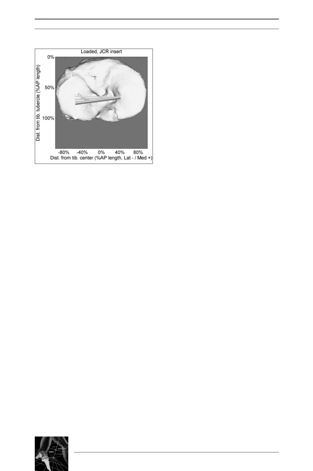
Fig. 3 : Loaded contact points Journey CR
REFERENCES
[1] BANKS S, BELLEMANS J, NOZAKI H
et al.
Knee
motions during maximum flexion in fixed and mobile-bea-
ring arthroplasties.
Clin Orthop 2003; 410: 131.
[2] AMIS AA, BULLAM, GUPTE CM
et al.
Biomechanics
of the PCL and related structures: posterolateral, postero-
medial and meniscofemoral ligaments.
Knee Surg Sports
Traumatol Arthrosc 2003; 11: 271-81.
[3] CHRISTEN B, HEESTERBEEK P, WYMENGA A
et
al.
Posterior cruciate ligament balancing in total knee repla-
cement: the quantitative relationship between tightness of
the flexion gap and tibial translation.
J Bone Joint Surg Br
2007; 89-B: 1046.
[4] PAGNANO MW, HANSSEN AD, LEWALLEN DG
et
al.
Flexion instability after primary posterior cruciate retai-
ning total knee arthroplasty.
Clin Orthop 1998; 356: 39.
[5] ROMERO J, STAHELIN T, BINKERT
et al.
The clini-
cal consequences of flexion gap asymmetry in total knee
arthroplasty.
J Arthroplasty 2007; 22-235-40.
[6] FREEMAN MA, PINSKEROVA V. The movement of
the knee studied by magntetic resonance imaging.
Clin
Orthop 2003; 35-43.
[7] JONG DE RJ, HEESTERBEEK PJC, WYMENGAAB.
A new measurement technique for the tibiofemoral contact
point in normal knees and knees with TKR.
Knee Surg
Sports Traumatol Arthroscop 2010; 18: 388-93.
[8] VICTOR J, VAN GLABBEEK F, VANDER SLOTEN J
et al.
An experimental model for kinematic analysis of the
knee.
J Bone and Joint Surg Am 2009 suppl 6: 150-63.