
Deepening Trochleoplasty: the Lyon Procedure
211
proximally into the muscle belly. After
inspection, the patella is retracted laterally,
providing adequate trochlear exposure.
Before starting the osseous procedure, the
synovium must be removed from the edge of
the trochlea. It is incised with a scalpel on the
cartilage edge and a periosteal elevator is used
to retract it away from the trochlear edge.
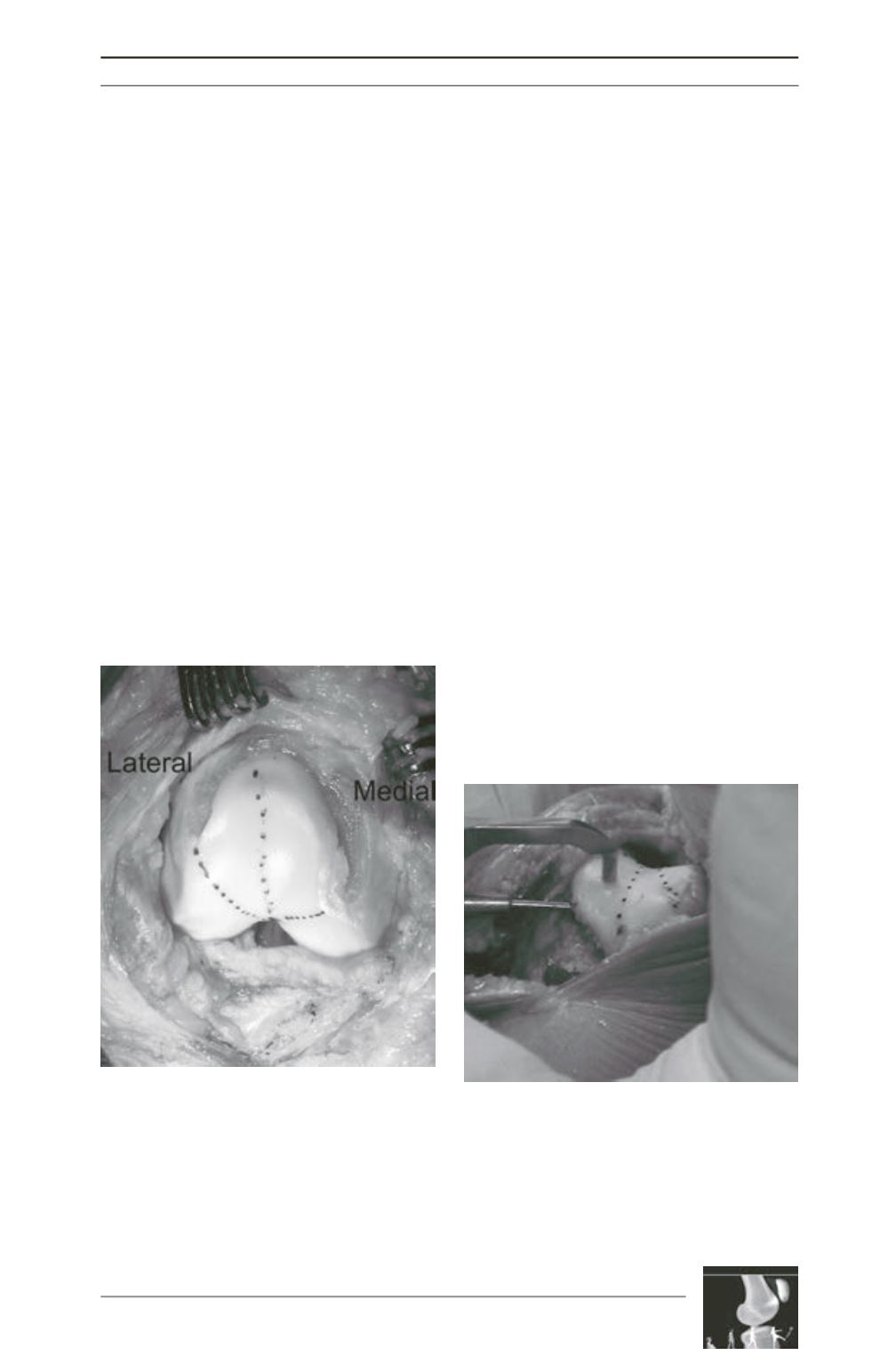
Trochlear planning
A line deviating 3 to 6 degrees laterally is
drawn proximally from the intercondylar notch
representing the bottom of the new groove. The
lateral margins are also drawn from the
intercondylar notch through the
sulcus
terminalis
of each condyle. The proximal part
of the new groove can be positioned according
to the TT-TG value in order to correct a possible
malalignment (fig. 2).
Bone removal
A strip of cortical bone (corresponding to the
amount of the bump) from the anterior femoral
cortex till the cartilage edge must be removed,
using a thin osteotome. This creates the access
to the under surface of the trochlea and
constitutes the first step to bring the new
trochlea to a normal position (not prominent)
and eliminate the bump.
The cancellous bone that lies under the trochlear
cartilage must also be removed to reshape and
reposition the new trochlea. Osteotomes and
curettes are helpful in the procedure, but a drill
with a specific depth guide is used for this
purpose. This depth guide is set to allow bone
removal from the cartilage undersurface
producing a uniform cartilage-bone shell of 5
millimeters. It avoids cartilage damage and
thermal necrosis which could result from
excessive bone removal (fig. 3). The shell
produced must be sufficiently compliant to
allow modeling. Without the guide, bone must
be removed especially from where the new
planned sulcus will lie. All bone that projects
beyond the anterior femoral cortex should be
removed (fig. 4).
Fig. 2 : The new trochlea is planned over the native
one. The central line deviating slightly laterally
represents the bottom of the groove. Note that
cortical bone has already been removed from the
osteochondral edge.
Fig. 3 : Cancellous bone removal with a drill and a
specific guide.


