
P.R.F. Saggin, P.G. Ntagiopoulos, P. Ferrua, D. Dejour
212
The cartilage-bone flap should sit flush on the
underlying bone bed. The bone bed should be
deepened in its central portion to recreate and
adequate the groove. Once the flap is
adequately modeled over the bone bed and the
trochlear conformation is satisfactory, fixation
is performed.
Fixation
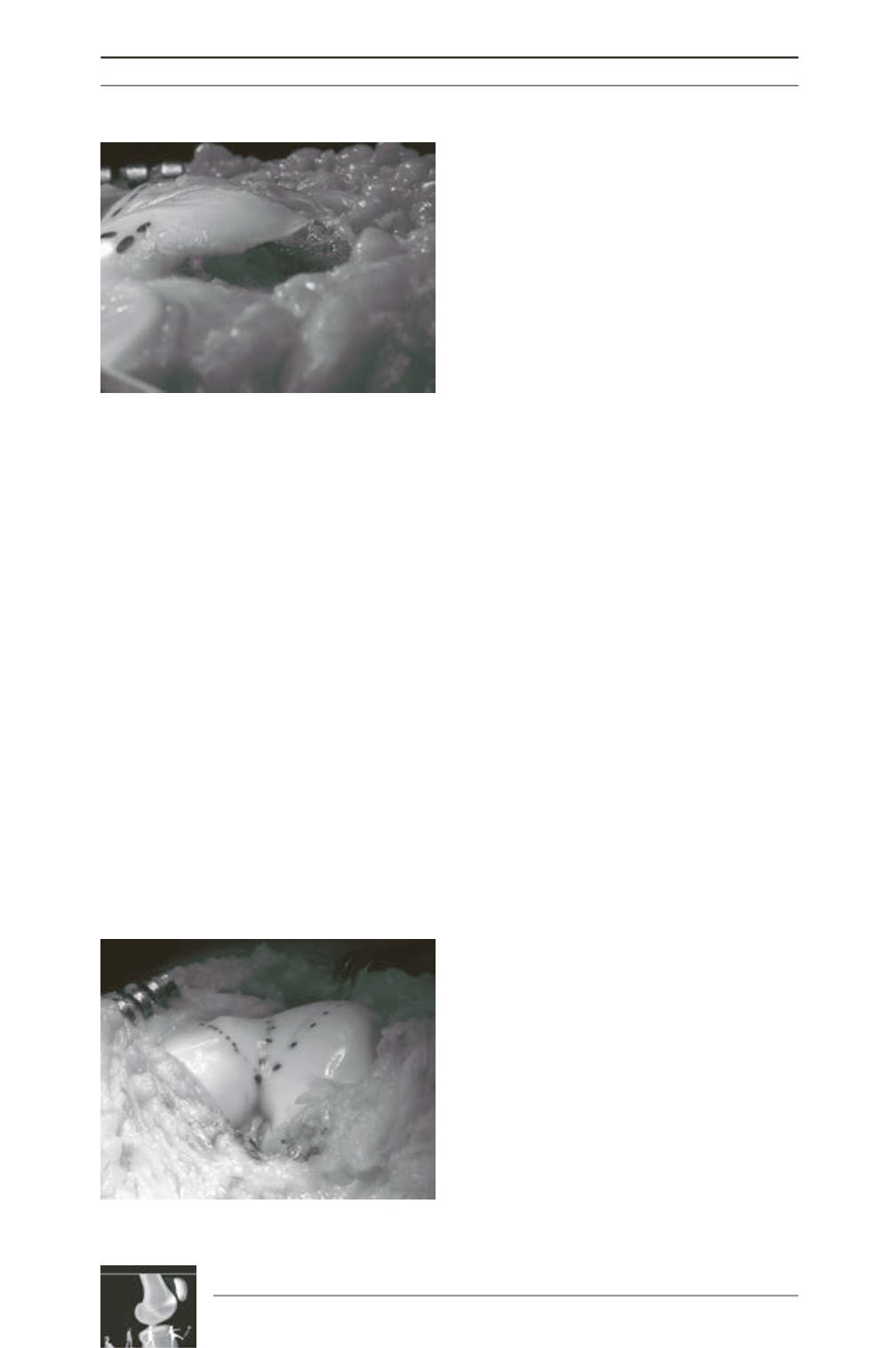
Absorbable vycril sutures are used to fix the
cartilage-bone flap to the underlying bone bed.
One suture is passed from each facet and tied
over the respective medial and lateral gutters;
this allows pulling down the new groove with
similar pressure of both facets on the cancellous
bone, promoting a perfect healing (fig. 5).
The synovium that was formerly dissected
away from the osteochondral margin is sutured
back to it. This protects the patellar cartilage
from the femoral bone and minimizes blood
loss through the exposed cancellous bone.
Patellar tracking check-up:
After satisfactory
trochlear shape is achieved, the associated
planned procedures are performed. We
routinely perform an associated medial
patellofemoral ligament reconstruction. After
the complete procedure is done, patellar
tracking and stability are checked.
Closure
Closure of the medial retinaculum is the final
step. No drains are installed.
Post-op protocol
Immobilization or weight restriction are not
necessary. Early mobilization on a continuous
passive motion device improves cartilage
nutrition and helps further trochlear modeling
by the patella. Immediate weight bearing is
allowed with an extension brace and flexion
must be regained without forced or painful
postures. Early rehabilitation goals are pain
and edema reduction and range of motion
recovery. The brace is removed when the
quadriceps strength allows the patient to walk.
Quadriceps strengthening with weights on the
foot or the tibial tubercle is prohibited in the
initial phase. After 45 days, cycling with weak
resistance and weight bearing proprioceptive
exercises may be initiated. From the 4
th
to the
6
th
month, running can be reinitiated and
quadriceps reinforcement with open kinetic
chain exercises between 0 and 60 degrees and
minor loads are allowed. Sports return is
allowed after the 6
th
month.
Control X-rays are obtained postoperatively
immediately and at 6 weeks. Six months after
the procedure, a CT scan is performed to
document the obtained correction.
Fig. 4 : The bone from the undersurface of the
trochlea has been removed. All bone that projects
beyond the anterior cortex of the femur must be
resected in order to eliminate the bump.
Fig. 5 : After fixation, trochlear anatomy resembles
that of normal knees.


